Bilateral VATS Surgery for Thymoma: Advanced Minimally Invasive Mediastinal Mass Removal
Minimally Invasive Thymoma Surgery with Faster Recovery
Introduction
Bilateral VATS Surgery for Thymoma is an advanced minimally invasive technique used for safe and effective mediastinal mass removal. At Naman Cancer Clinic & Research Centre, Dr. Priyansh Jain successfully treated a young patient with thymoma using bilateral VATS surgery, achieving complete tumour removal, faster recovery, and excellent postoperative outcomes.
A mediastinal mass is an abnormal growth located in the mediastinum — the central compartment of the chest situated between the lungs. Among the various mediastinal tumours, thymoma is a relatively rare tumour arising from the thymus gland. Early diagnosis and timely surgical management play a crucial role in achieving excellent outcomes.
At Naman Cancer Clinic & Research Centre, we recently managed a challenging case of thymoma in a young patient using a minimally invasive bilateral VATS (Video-Assisted Thoracoscopic Surgery) approach.
Case Summary
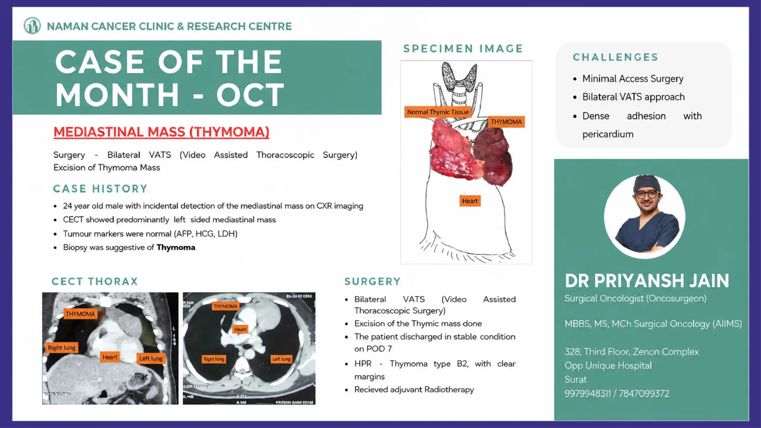
A 24-year-old male patient was incidentally diagnosed with a mediastinal mass during routine chest X-ray (CXR) imaging. The patient did not have major symptoms, highlighting how some chest tumours may remain silent and get detected accidentally during investigations performed for unrelated reasons.
Further evaluation with Contrast Enhanced CT (CECT) Thorax revealed a predominantly left-sided mediastinal mass. Blood tumour markers including AFP, HCG, and LDH were within normal limits, helping rule out certain germ cell tumours.
A biopsy of the lesion suggested the diagnosis of thymoma.
Understanding Thymoma
The thymus is a small organ located in the upper chest behind the sternum and plays an important role in immunity during childhood. Tumours arising from this gland are known as thymomas.
Although thymomas are uncommon, they are the most frequent tumours of the anterior mediastinum in adults. Some patients may present with symptoms like:
- Chest pain
- Cough
- Breathing difficulty
- Myasthenia gravis (muscle weakness disorder)
However, many patients remain asymptomatic and are diagnosed incidentally, as seen in this case.
Imaging Findings
CECT Thorax demonstrated a well-defined mediastinal mass located predominantly on the left side of the chest cavity. The tumour was closely related to surrounding structures, including the pericardium (covering of the heart), making surgical planning crucial.
Radiological imaging helped assess:
- Tumour size and extent
- Relationship with nearby vital structures
- Feasibility of minimally invasive surgery
Surgical Challenges
Managing mediastinal tumours can be technically demanding because of the presence of vital structures such as:
- Heart
- Great vessels
- Lungs
- Pericardium
- Phrenic nerves
In this patient, the major surgical challenges included:
- Performing surgery through a minimally invasive approach
- Requirement of bilateral VATS access
- Dense adhesions between the tumour and pericardium
Despite these challenges, a minimally invasive strategy was chosen to reduce postoperative pain, improve recovery, and minimize hospital stay.
Bilateral VATS Surgery for Thymoma
The patient underwent Bilateral VATS (Video-Assisted Thoracoscopic Surgery) excision of the thymoma.
VATS is an advanced minimally invasive surgical technique performed using small incisions and a thoracoscopic camera. Compared to traditional open chest surgery, VATS offers several advantages:
- Smaller incisions
- Less postoperative pain
- Faster recovery
- Reduced blood loss
- Better cosmetic outcome
- Shorter hospital stay
Using bilateral thoracoscopic access allowed complete visualization and safe removal of the thymic tumour.
Histopathology Report
Final Histopathological Report (HPR) confirmed:
- Thymoma Type B2
- Clear surgical margins
Achieving clear margins is one of the most important goals in cancer surgery, as it significantly reduces the risk of recurrence.
Considering the tumour characteristics, the patient subsequently received adjuvant radiotherapy for optimal long-term disease control.
Recovery and Outcome
The postoperative recovery was smooth, and the patient was discharged in stable condition on postoperative day 7.
This case highlights how advanced minimally invasive oncological surgery can successfully manage even complex mediastinal tumours with excellent outcomes.
Key Learning Points
- Thymoma may remain asymptomatic and get detected incidentally.
- Proper imaging and biopsy are essential for diagnosis and surgical planning.
- Minimally invasive Bilateral VATS surgery is feasible in selected mediastinal tumours.
- Complete tumour excision with clear margins remains the cornerstone of treatment.
- Multidisciplinary management including surgery and radiotherapy improves outcomes.
Conclusion
Thymoma is a rare but treatable mediastinal tumour when diagnosed early and managed appropriately. Advances in minimally invasive thoracic oncology surgery now allow safe and effective tumour removal with faster patient recovery.
At Naman Cancer Clinic & Research Centre, we continue to focus on evidence-based cancer care with advanced surgical techniques aimed at improving patient outcomes and quality of life.
Also read,
Dr Priyansh Jain
Surgical Oncologist (Oncosurgeon)
Naman Cancer Clinic & Research Centre, Surat
Also read: Advanced Oral Cancer Surgery with ALT Free Flap Reconstruction


